FILOVIRIDAE
Enveloped, non-segmented,(-) ssRNA Group V
Structure
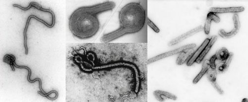
Display unusual variability in shape.
Virions consist of an envelope, a nucleocapsid, a polymerase complex, and a matrix protein.
Capsid/nucleocapsid is elongated and exhibits helical symmetry.
Capsids are enveloped.
Virions are filamentous, or pleomorphic, flexible with extensive branching.
Fishhook, U- or 6-shaped and circular forms occur particularly after purification.
800-970 nm (sometimes 4,000 nm) in length x 80 nm diameter.
Surface projections are spaced widely apart, distinctive knob-shaped peplomers that cover evenly the surface and are embedded in a lipid bilayer.
Surface projections are composed of one type of glycoproteins (GP).
GPs (spikes or peplomers or surface projections) are 10 nm long and spaced 10 nm apart.
Source: ICTV
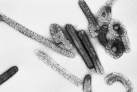
Source: CDC/ Dr. Erskine Palmer, Russell Regnery
This negative stained transmission electron micrograph (TEM) depicts a number of filamentous Marburg virions, which had been cultured on Vero cell cultures, and purified on sucrose, rate-zonal gradients. Note the virus’s morphologic appearance with its characteristic “Shepherd’s Crook” shape. Magnified approximately 100,000x.
Source: CDC
Taxonomy
The family Filoviridae (Filum is Latin for 'thread') constitutes, together with the families Paramyxoviridae and Rhabdoviridae, the order Mononegavirales.
Within the family there is a single genus, Filovirus, and a separation into two sero-/genotypes, Marburg and Ebola.
Marburg
Ebola
Zaire
Infects humans
Sudan
Infects humans
Tai or Ivory Coast
First Ebola virus known to be passed to humans from other animals) - naturally infected chimpanzees of the Tai Forest in the Ivory Coast.
Reston
Isolated from infected monkeys delivered to Reston (Virginia) in 1989.
Appears to be spread only among monkeys and not humans.
Fatal in monkeys.
Source: University of Cape Town
Genome
The genome is not segmented and contains a single molecule of linear negative-sense, ssRNA.
The complete genome is 18-19 kb long.
The 5'-end of the negative-sense strand does not have a covalently attached terminal protein; genome does not have cap.
The 3'-terminus has conserved nucleotide sequences (leader), in genera of same family and has no poly (A) tract.
The viral genome encodes structural proteins and non-structural proteins.
Virions consist of 5 structural protein(s) located in the envelope (surface glycoprotein), nucleocapsid (NP), polymerase complex (transcriptase-polymerase component and RNA-dependent RNA transcriptase-polymerase), and a matrix protein.
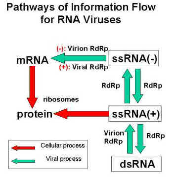
The viral RNA dependent RNA polymerase binds the encapsidated genome at the leader region, then sequentially transcribes each genes by recognizing start and stop signals flanking viral genes.
mRNAs are capped and polyadenylated by the L protein during synthesis.

Replication
Filovirus replication strategies are not completely understood.
The replication of Filoviruses resembles that of other (-) sense RNA viruses.
Virus attaches to host receptors through G glycoprotein and is endocytosed into vesicles in the host cell.
Since virion RNA is not readily detected in the cells, a fast replication process most likely occurs with (-) sense RNA being released rapidly within virions.
Filovirus transcription and replication are mediated by a single virus-encoded polymerase in the cytoplasm of the infected cell.
The negative-sense RNA genome is transcribed into monocystronic, polyadenylated subgenomic RNAs which are translated into seven structural proteins.
Sequential transcription, viral mRNAs are capped and polyadenylated in the cytoplasm.
(+) sense antigenome serves as a template for (-) sense progeny genomes.
Particles mature at the plasma membrane.
Buds from plasma membrane.
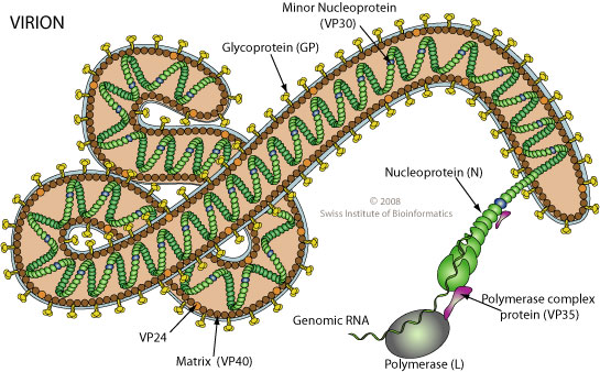
Source: Swiss Institute of Bioinformatics via viralzone.
Diseases
Virus primarily targets primates and also guinea pigs, hamsters, and mice.
These viruses cause viral hemorrhagic fevers, characterized by bleeding and coagulation abnormalities, often leading to death.
Filoviruses are classified as "Biological Level 4" agents (WHO; Risk Group 4) based on their high mortality rate, person-to-person transmission, potential for aerosol infectivity, and absence of vaccines and chemotherapy.
Marburg
This zoonotic virus was first recognized in 1967, when outbreaks of hemorrhagic fever occurred simultaneously in laboratories in Marburg and Frankfurt, Germany and in Belgrade, Yugoslavia (now Serbia).
31 lab workers in Marburg were exposed to tissues and blood from African green monkeys (Cercopithecus aethiops) imported from Uganda and infected with Marburg virus; 7 died.
Sporadic cases of Marburg hemorrhagic fever in humans have occurred in Kenya and Zimbabwe.
Nosocomial cases have been reported with a mortality of about 25%.
Ebola (humans)
First recognized in Zaire and Sudan in 1976.
Subtypes: Sudan (mortality 51%), Zaire (mortality 88%).
Death rate ranges between 50 to 90% according to subtype.
About 20% of the population of rural areas in Central Africa has antibodies to Ebola.
Reservoirs
Still not known.
Virus isolated directly from monkeys and from laboratory inoculation of guinea pigs.
Transmission and Control
The mechanisms through which filoviruses spread are not fully understood.
The route of transmission from animals to humans is unknown.
Person-to-person transmission occurs primarily through physical contact with infected bodily fluids, such as semen or infected blood or vomit.
By close contact with infected individuals and exposure to contaminated needles and syringes.
Monkeys caught in the wild are an important source for the introduction of filoviruses.
Quarantine of imported non-human primates and professional handling of animals will help prevent introduction into humans.
Because of the biohazardous nature of the agents, recombinant vaccines would be an attractive approach in the future.
Immunization of monkeys with purified NP and GP has demonstrated the induction of the humoral and cellular immune responses and protection of animals against challenge with lethal doses.
No effective vaccine today.