HEPADNAVIRIDAE
An enveloped virus.
Group VII - ds/ss DNA encodes reverse transcriptase (as Caulimoviridae) - a 'pararetrovirus'.
Hepatitis virus B (HVB) is the single most important cause of cancer in humans.
Excellent progress in anti-HBV vaccines.
Most hepadnaviruses will only replicate in specific hosts, and this makes experiments using in vitro
methods very difficult.
HBV still can not be grown in culture.
Only the woodchuck and the duck hepatitis viruses have been recently grown in culture.
The virus resists drying for up to 1 week.
Structure
Virions are spherical to pleomorphic.
Filamentous virions occur when HBsAg (hepatitis B surface antigen S) is lacking.
40 - 48 nm in diameter, with no evident surface projections.
Capsid diameter is 35 nm and is made of 180 capsomers.
Lipids are present in the envelope as phospholipids, sterols, and fatty acids.
The virion is not a simple naked icosahedral structure, as are those of all the other viruses with small genomes, but rather has a nucleocapsid (core) surrounded by a very tight envelope
containing three viral glycoproteins.
The major protein of the capsid is called the HBV core antigen (HBcAg).
The external glycoproteins are called HBV surface antigens or HBsAg.
Virion envelope proteins consist of three antigenically distinct species (S, M and L proteins).
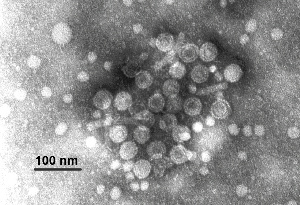
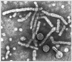
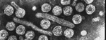
Electron micrographs of circular and filamentous forms of HBV. Source: ICVT website.
Genome
Monopartite (non-fragmented) circular DNA.
Very small linear genomes consisting of a (-) sense strand of
3.2 kb and a (+) sense incomplete
strand of 1.7-2.8 kb.
One DNA strand, the minus strand, has a protein covalently attached to the 5' end.
The other strand has an RNA oligonucleotide (small molecule) attached at its 5' end.
The virus carries its own RNA-dependent DNA polymerase (i.e. reverse transcriptase).
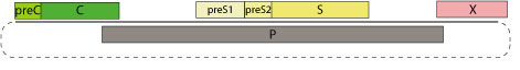
There are four known genes in the virus (C, P, S, X) that encodes 7 polypeptides:
C encodes the core protein (structural)
P encodes DNA polymerase, RT, and RNase H activities
S encodes 3 polypeptides (surface antigens) - (also called HBsAg)
X encodes a small transactivator of viral transcription
Synthesis of the viral protein is complex.
The viral genome is very compact.
Over half of it is translated in two reading frames.
The P gene requires about three-quarters of the coding capacity
of the genome
and this gene overlaps each of the other three genes (see figure
below).
The S gene is completely contained within the P gene.

Virion DNA is encapsulated in core protein, which is surrounded
by a lipid envelope coat containing
the S (surface) antigen.
HBsAg (hepatitis B surface antigen S) is produced in large quantities and it found in serum.
Antibody to S antigen provides protective immunity and is the keystone of the immunization policy.
Current vaccination schedule calls for 3 injections of S protein over a period of 6 months.
Cycle of Infection
Tropism to liver cells (hepatocytes)
Attachment/penetration events: not known
Attaches to susceptible cell through recognition of a receptor still not identified
Mechanism of viral uptake is also unknown
Transport to the nucleus depends on the phosphorylation of the
capsid protein and is
mediated by cellullar transport receptors.
Maturation/release: assembly in cytoplasm, release mechanism is not known yet
May transform permissive host cells
Released from cell by exocytosis
Viral Replication
They have a unique and complex mode of replication, because they replicate through an RNA intermediate (which they translate back into DNA using reverse transcriptase).
Circular DNA is found soon after infection in the nucleus of the host cell.
Genome converted into a form called covalently closed circular DNA (cccDNA).
The resulting cccDNA does not integrate into the host genome nor does it replicate as an episome.
RNA transcripts are capped and polyadenylated.
A polymerase (RT) converts RNA to DNA in the particles as they form in the cytoplasm.
Budding is through internal membranes.
Integration of viral DNA into the cellular genome takes place infrequently and is therefore unlikely to be essential for replication.
Integration with cellular DNA is highly significant to the proposed involvement of HBV with liver cancer.
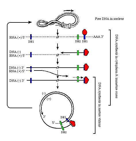
The following images describe the replication process of the virus:
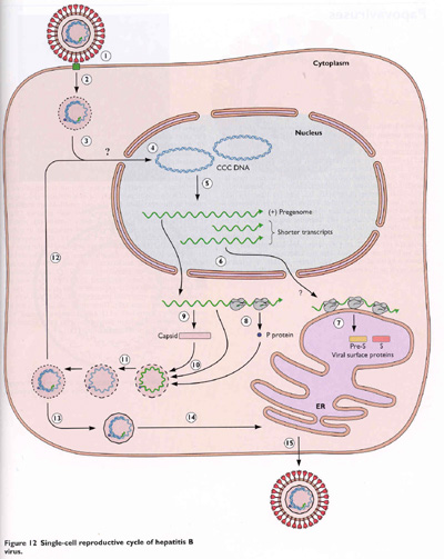
Source: Flint el al. 2000. Principles of Virology. ASM.
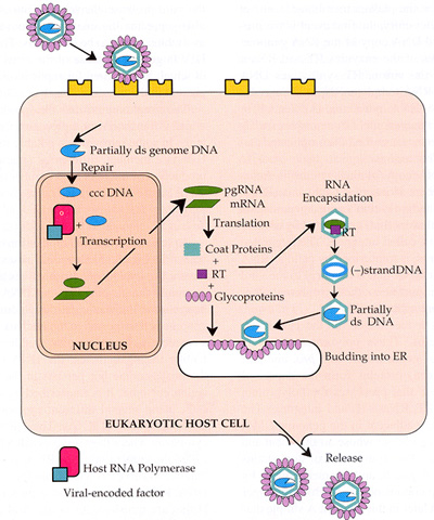
Source: Strauss & Strauss, 2008, Virus and Human Disease. Academic Press.
Human Pathogens
Orthohepadnavirus
Hepatitis B virus (HBV) -- acute and chronic hepatitis.
200,000 cases reported annually in the United States.
In 10% of the patients, the infection is not cleared and becomes
chronic; and, can be
asymptomatic leading to cyrrhosis and death.
There are approximately 1.2 million carriers of HBV in the US, and 350 million worldwide.
Up to 90% of neonates become chronically infected due to immature
state of the
immune system.
HBV could be linked to hepatocellullar carcinoma (500,000 deaths worldwide a year)
Because chronic infection of neonates is a major reservoir of the virus in nature, elimination of this source of the virus could lead to eradication of the virus in the human population.
Animal Pathogens
Orthohepadnavirus
Woodchuck hepatitis virus
Ground squirrel hepatitis virus
Red-bellied squirrel hepatitis virus
Old and new world primates viruses
Wooly monkeys, orangutans, gorillas, gibbons, and chimpanzees.
Avihepadnavirus
Pekin duck hepatitis virus
Heron hepatitis virus
Snow geese hepatitis virus
The Hepatitis
Delta Agent (HDAg)
May have arisen from viroids that evolved a mechanism for packaging using a helper virus.
Hepatitis Delta Agent is not a member of Hepadnaviridae.
It causes hepatitis D (delta).
Its genome is unrelated to the genomes of hepadnaviruses.
It works along with hepatitis B to cause infection.
A satellite of hepatitis B virus, hepatitis delta agent is a small
circular RNA virus which requires the
help of a hepadnavirus like HBV for its own replication.
HDV is not a defective interfering particle, but rather a satellite virus, a natural subviral satellite of HBV.
HDV is replication defective.
It requires the surface antigen of HBV for the encapsidation of
its own genome.
The mRNA for HDAg is exported to the cytoplasm and translated
into a protein of 195 amino acids,
referred to as the small delta antigen or S-HDAg. This protein
is required for RNA replication.
The envelope proteins on the outer surface of HDV are entirely provided by HBV.
HDV is extremely prolific.
The serum of an infected individual can contain up to 1012 (1 trillion) RNA-containing HDV particles per milliliter.
The only known natural hosts for HDV are humans.
It has been experimentally transmitted to chimpanzees and woodchucks in co-infection with HBV.
HDV is present worldwide and in all age groups.