Differences in the Multiplication of Phages and Animal Viruses
Follows the basic pattern of bacteriophage multiplication but has some differences:
Mechanism of entering the host cell.
Once the virus is inside, the synthesis and assembly of the new viral components are somewhatdifferent, partly because of the differences between procaryotic and eucaryotic cells.
Animal viruses may have certain types of enzymes not found in phages.
Mechanisms of release, and the effects on the host cell are also different.
Animal viruses do not possess appendages like the tail fibers of T4.
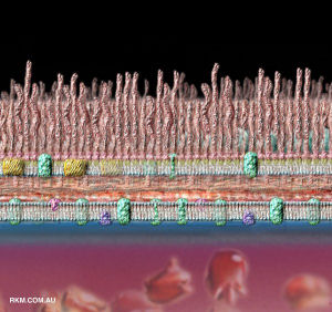 Cell wall of G- bacteria |
 Cell wall of G+ bacteria |
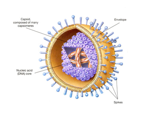
A typical animal virus
Steps in the Multiplication of Animal Viruses
Attachment
- Similar to that of phages: complementary receptor sites on the host's cell surface.
- Receptor sites of animal cells are proteins and glycoproteins of the plasma membrane.
- The attachment sites of animal viruses are distributed over the surface of the virus.
- The sites themselves vary from one group of viruses to another.
- In adenoviruses, the attachment sites are small fibers at the corners of the icosahedron.
- In many of the enveloped viruses, such as influenza virus, the sites are spikes located on the surface of the envelope.
- As soon as one spike attaches to a host receptor, additional receptor sites on the same cell migrate to the virus.
- Attachment is completed when many sites are bound.
- Receptor sites are inherited characteristics of the host.
- The receptor for a particular virus can vary from person to person which accounts for the individual differencesin susceptibility to a particular virus.
- Understanding the nature of attachment can lead to the development of drugs that prevent viral infections.
- Monoclonal antibodies that combine with a virus's receptor site or the cell's receptor site may soon be used to treat some viral infections.
- One experimental treatment for AIDS involves injections of receptor sites.
- If HIV attaches to these, it cannot attach to natural receptor sites on cells.
Penetration
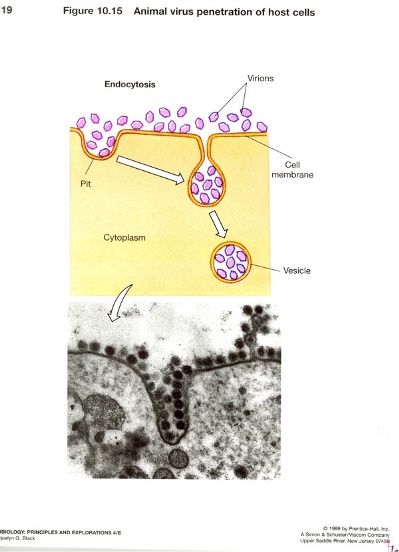
- Occurs by endocytosis, an active cellular process by which nutrients and other molecules are brought into a cell.
- A cell's membrane continuously folds inward to form vesicles.
- Vesicles contain elements that originate outside the cell and are brought into the interior of the cell to be digested.
- If a virion attaches to a small out-folding (microvillus) on the membrane of a potential host cell, the cell willenfold the virion into a fold of plasma membrane, forming a vesicle.
- Once the virion is enclosed within the vesicle, its viral envelope is destroyed.
- The capsid is digested when the cell attempts to digest the vesicle's contents, or the non-enveloped capsid may be released into the cytoplasm of the host cell.
- Enveloped viruses can penetrate by an alternative method called fusion, in which the viral envelope fuseswith the membrane and releases the capsid into the cell's cytoplasm.
- HIV and herpesviruses penetrate the cells by fusion.
- In bacteriophages, viral DNA is injected into the host cell, there is no endocytosis or fusion.
Uncoating
- The separation of the viral nucleic acid from its protein coat. Poorly understood process that varies with viral type.
- Some animal viruses accomplish uncoating by the action of lysosomal enzymes contained inside the phagocytic vacuoles and coated vesicles of the host cell.
- These enzymes degrade the proteins of the viral capsid.
- The uncoating of poxviruses is completed by a specific enzyme encoded by the viral DNA and synthesized soon after infection.
- For other viruses, uncoating appears to be exclusively caused by enzymes in the host cell cytoplasm.
- For poliovirus, uncoating seems to begin while the virus is still attached to the host cell's plasma membrane.
- Uncoating is not required in bacteriophages.
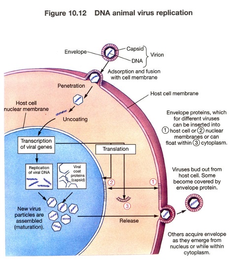
The Biosynthesis of DNA Viruses
- Generally, DNA-containing viruses replicate their DNA in the nucleus of the host cell by using viral enzymes.
- DNA viruses synthesize their capsid and other proteins in the cytoplasm by using host cell enzymes.
- Then proteins migrate into the nucleus and are joined with the newly
synthesized DNA to form virions.
- Virions are transported along the endoplasmatic reticulum into the host cell's membrane for release.Examples: herpes, papova , adeno and hepadnaviruses.
- Poxviruses are an exception because all of their components are synthesized in the cytoplasm.
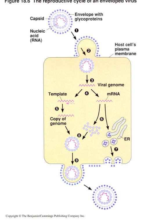
The Reproductive Cycle of an Enveloped Virus
Budding Mechanism
Patterns of Biosynthesis of DNA Viruses
Use the nucleus of the host cell to reproduce their nucleic acids.
Main Cases:
Adenoviridae (dsDNA)
Cellular transcriptase transcribes viral DNA in nucleus.
Poxviridae (dsDNA)
Viral multiplication is started by viral transcriptase; the viral components are synthesized and assembled in the cytoplasm of the host cell.
Viral enzyme transcribes viral DNA in cytoplasm.
Poxviruses contain their own transcriptase (RNAP), others used the host cell's transcriptase
Herpesviridae (dsDNA)
Cellular transcriptase transcribe viral DNA in nucleus. Prophage stays dormant in episome in cytoplasm.
Papovaviridae (dsDNA)
Some types of Papillomavirus are capable of transforming cells and causing cervical cancer. Nobel prize 2008.
Viral DNA is replicated in the host cell's nucleus along with host cell chromosomes.
Transcription uses host transcriptase (RNA polymerase).
Capsid proteins migrate to nucleus, maturation occurs, then after assembly of complete viruses they are released.
Hepadnaviridae (DNA, RT)
Vrus causes hepatitis B.
Hepatitis A, C, D, E, F, and G are caused by non-related ssRNA viruses.
Differ from other DNA viruses because they synthesize DNA by copying RNA, using viral reverse transcriptase (RT).
Cellular enzyme transcribes viral DNA in nucleus.
RT copies mRNA to make viral DNA in nucleus.
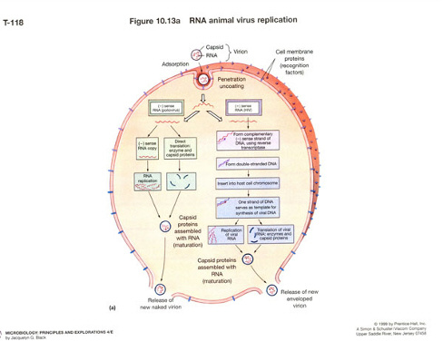
Pattern of Biosynthesis of RNA Viruses
Same as that of DNA viruses, except that several different mechanisms of mRNA formation occuramong the four different groups of RNA viruses.
RNA viruses multiply in the host cell's cytoplasm.
The major differences among the multiplication processes of these viruses lie in how mRNA and viral RNA are produced.
Once viral RNA and viral proteins are synthesized, maturation occurs by similar means among all animals viruses.
Main Cases:
Picornaviridae (ss+RNA)
Includes the poliovirus one of the smallest viruses.
As it has a sense strand (or + strand) it acts as mRNA.
After attachment, penetration, and uncoating is completed, the ss viral (+)RNA is translated into two principal proteins, which inhibit the host cell's synthesis of RNA and protein, and which forms an enzyme called RNA-dependent RNA polymerase.
This enzyme catalyzes the synthesis of another strand of (-)RNA, which is complementary in basesequence to the original infecting strand.
The new strand is called 'antisense or - strand', and serves as a template to produce additional (+) strands.
The (+) strands may serve as mRNA for the translation of capsid proteins, may become incorporated into capsid proteins to form a new virus, or may serve as a template for continued mRNA multiplication.
Once viral (+)RNA and viral protein are synthesized, maturation and further exit occurs.
Togaviridae (ss +RNA)
Includes arthropod-borne alphaviruses or arboviruses.
Only family with an unusual icosaderal envelope.
After a (-) strand is made from the (+)strand, two types of mRNA are transcribed from the (-) strand.
One type of mRNA is a short strand that codes for envelope proteins; the other, longer strand serves as mRNA for the synthesis of capsid proteins..
Rhabdoviridae (ss -RNA)
Includes the genus Lyssavirus, bullet-shaped virus that causes rabies.
Contain an RNA-dependent RNA polymerase that uses the (-) strand as a template from which toproduce a (+) strand.
The (+)strand serves as mRNA and as a template for synthesis of new viral (-)RNA.
Viral enzyme copies viral (-)RNA to make mRNA in cytoplasm.
Reoviridae (dsRNA)
Found in respiratory and enteric systems of humans.
Not associated with any diseases when discovered.
They were considered orphans (unclaissified)
Reo: respiratory, enteric, orphan.
Capsid is digested upon entering a host cell.
Viral enzyme copies (-)strand RNA to make mRNA in the cytoplasm, where it is used to synthesize more viral proteins.
One of the newly synthesized viral proteins acts a RNA-dependent RNA polymerase to produce more (-) strands of RNA.
The RNA (+) and (-) strands form the dsRNA that is then surrounded by capsid proteins.
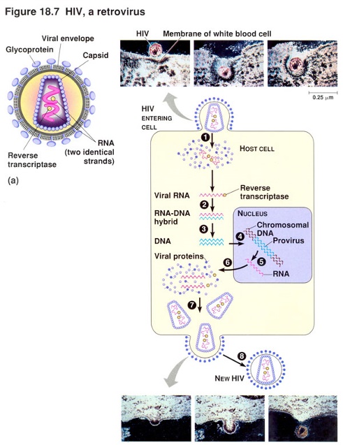
Retroviridae (ssRNA, RT)
Many infect vertebrates.
The genus Lentivirus includes types HIV1 and HIV2.
Others types may cause cancer.
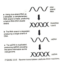
Carry their own polymerase knowns as RNA-dependent DNA polymerase.
RT carries out a reaction (RNA to DNA) that is exactly the reverse of the familiar transcription of DNA to RNA.
RT uses the RNA of the virus to synthesize a complementary strand of DNA, which in turn is replicatedto form dsDNA.
RT degrades the original viral RNA.
The formation of complete viruses requires that DNA be transcribed back into the RNA that will serveas mRNA for viral protein synthesis and be incorporated into new virions.
Before transcription can take place, the viral DNA must be integrated into the DNA of a host cell chromosome.
In this integrated state (lysogeny), the viral DNA is called a provirus.
Unlike a prophage, the provirus never comes out of the chromosome.
Once the provirus is integrated into the host cell's DNA, several things can happen.
Sometimes the provirus simply remains in a latent state and replicates when the DNA of the host cell replicates.
In other cases, the provirus becomes transcribed.
Or, produces new viruses, which may infect adjacent cells.
The provirus can also convert the host cell into a tumor cell.
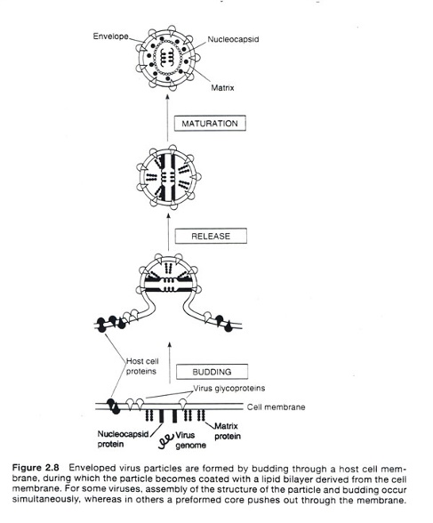
Maturation and Release
- The first step in viral maturation is the assembly of the protein capsid.
- It is a spontaneous process.
- The capsids of many of the RNA-containing animal viruses are enclosed by an enveloped consisting of protein, lipid, and carbohydrate (e.g. orthomyxoviruses and paramyxoviruses).
- The envelope protein is synthesized by the virus and is incorporated into the plasma membrane of the host cell.
- The envelope lipid and carbohydrate are synthesized by host cell enzymes and are present in the plasma membrane.
- The envelope actually develops around the capsid by a process called budding.
- After attachment, penetration, uncoating, and biosynthesis of viral nucleic acid and protein, the assembled capsid with nucleic acid pushes through the plasma membrane.
- As a result, a portion of the membrane, now the envelope, adheres to the virus.
- This extrusion of a virus from a host cell is one method of release.
- Budding does not immediately kill the host cell, and in some cases the host cell survives.
- Non-enveloped viruses are released through ruptures in the host cell plasma membrane.
- In contrast to budding, this type of release usually results in the death of the host cell.