MULTIPLICATION OF BACTERIOPHAGES
Although the means by which a virus enters and exists a host cell may vary, the basic mechanism of viral multiplication is similar for all viruses.
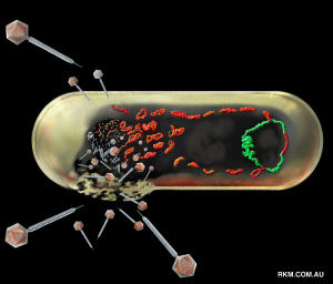
The best-understood viral life cycles are those of the T-even series (T2, T4, and T6) in Escherichia coli.
Two Alternative Mechanisms
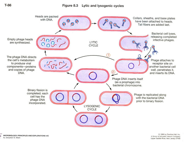
Lytic Cycle
The lytic cycle ends with the lysis and death of the host cell.
Lysogenic Cycle
The lysogenic cycle allows the cell to remain alive.
The T-even Bacteriophage
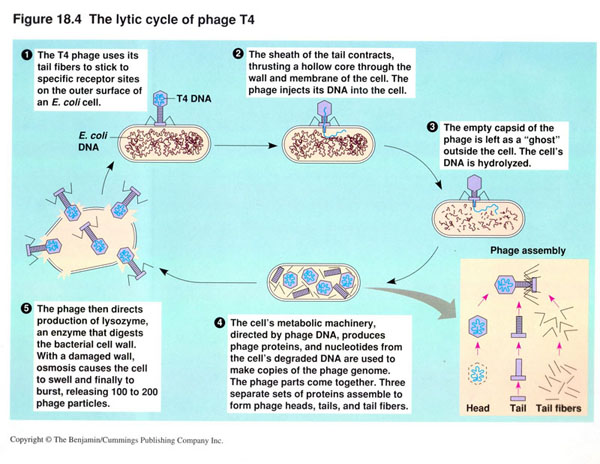
The Lytic Cycle
T-even virions are large, complex, and nonenveloped, with a characteristic head and tail structure.
The length of their DNA is about 6% of that contained in Escherichia coli, yet the phage has enough DNA for over 100 genes.
The multiplication cycle of these phages, like that of all viruses, occurs in five distinct stages.
Attachment (adsorption)
Penetration
Biosynthesis
Maturation (assembly)
Release
Attachment
Occurs after a chance collision between phage particles and bacteria.
An attachment site on the virus attaches to a complementary receptor site on the bacterial cell.
It is a chemical interaction in which weak bonds are formed between the attachment and receptor site molecules.
T-even bacteriophages use fibers at the end of the tail as attachment sites.
The complementary receptor sites are located on the bacterial cell wall.
T4 viruses on E. coli pilus
Penetration
Bacteriophage injects its DNA into the bacterium.
Bacteriophage 's tail releases an enzyme (lysozyme), which breaks down a portion of the bacterial cell wall.
The tail sheath of the phage contracts, and the tail core is driven through the cell wall.
When the tip of the core reaches the plasma membrane, the DNA from the bacteriophage's head passes through the tail core, through the plasma membrane, and enters the bacterial cell.
The capsid remains outside the bacterial cell.
The phage particle functions like a hypodermic syringe to inject its DNA into the host cell.
Biosynthesis
Once the bacteriophage DNA has reached the cytoplasm of the host cell, the biosynthesis of viral nucleic acid and protein occurs.
Host protein synthesis is stopped by virus-induced degradation of the host DNA, viral proteins interfere with transcription, or the repression of translation.
Phage uses host cell's nucleotides and several of its enzymes to synthesize many copies of phage DNA.
After synthesis of viral proteins begins, any RNA transcribed in the cell is mRNA transcribed from phage DNA for the biosynthesis of phage enzymes and capsid proteins.
Host cell's ribosomes, enzymes, and amino acids are used for translation.
Genetic controls regulate when different regions of phage DNA are transcribed into mRNA during the multiplication cycle.
Early messages are translated into early phage proteins, the enzymes used in the synthesis of phage DNA.
Late messages are translated into late phage proteins for the synthesis of capsid proteins.
The Eclipse Period
For several minutes following infection, complete infective phages cannot be found in the host cell.
Only separate components (DNA and protein) can be detected.
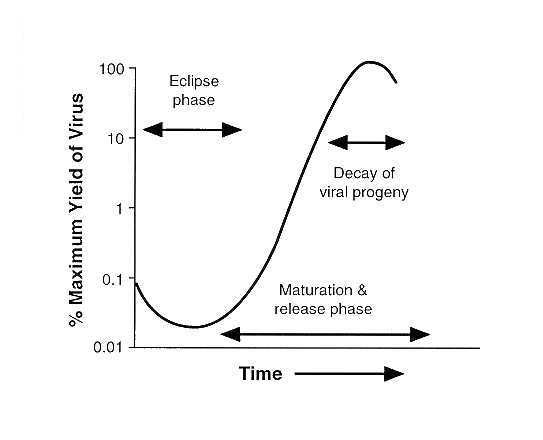
Maturation
Bacteriophage DNA and capsids are assembled into complete virions.The viral components essentially assemble into a viral particle spontaneously, eliminating the need for many non-structural genes and gene products.
The phage heads and tails are separately assembled from protein subunits, and the head is filled with phage DNA and attached to the cell.
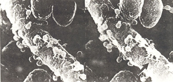
Release
Escape of virions from host cell due to lysis.
Lysis makes the plasma membrane break open (it lyses).
Lysozyme, which is coded for by a phage gene, is synthesized within the cell.
Lysozyme causes the bacterial cell wall to break down, and the newly produced bacteriophages are released from the cell.
The released bacteriophages infect other susceptible cells in the vicinity, and the viral multiplication cycle is repeated within those cells.
The time that elapses from attachment to release is known as the burst time and averages 20-40 minutes.
The number of newly synthesized phage particles released from a single cell is referred as burst size and usually ranges from about 50 to 200.
Lambda phage leaving an E. coli cell
The Lysogenic Cycle
In contrast to T-even bacteriophages, some viruses do not cause lysis and death of the host cell when they multiply.Some phage can either proceed through a lytic cycle or begin a lysogenic cycle by incorporating their DNA into the host's cell's DNA (lysogeny: phage remains latent or inactive).
Phages that can multiply by either mechanism are called lysogenic or temperate phages.
The participating bacterial host cells are called lysogenic cells.
The Lysogenic Cycle of Temperate Bacteriophage Lambda
Steps:
- Upon penetration into an Escherichia coli cell, the originally linear phage DNA forms a circle.
- This circle can multiply and transcribed, leading to the production of new phage and to cell lysis (the lytic cycle).
- Alternatively, the circle can recombine with and become part of the circular bacterial DNA (the lysogenic cycle).
- The inserted phage DNA is called a prophage.
- Every time the host cell's machinery replicates the bacterial chromosome, it also replicates the prophage DNA.
- The prophage remains latent or inactive within the progeny cells.
- A rare spontaneous event, or the action of UV light or certain chemicals, can lead to the excision (popping out) of the phage DNA, and to initiation of the lytic cycle.
Some Consequences of Lysogeny
- The lysogenic cells are immune to reinfection by the same phage.
- The host cell is not immune to infection by other phage types.
- The host cell may exhibit new properties:
- Corynebacterium diphtheriae only produces toxin when it carries a temperate phage that has the gene coding for the toxin.
- The toxin produced by Clostridium botulinum is encoded by a prophage gene, as is the cholera toxin produced by Vibrio cholerae.
- Scarlet fever results from a toxin released by lysogenized Staphylococcus aureus.
- Epsilon phage of Salmonella sp. alters surface lipopolysaccharides by altering activity of enzymes involved in their synthesis.
- Changes antigenic character of the surface and its susceptibility to specific antibody.
- Makes specialized transduction possible.
- Packaging of bacterial DNA along with its own DNA in the same capsid.
Animal Viruses can Undergo Processes very Similar to Lysogeny
They may remain latent for long periods without multiplying or remain inserted in the chromosome or separate in a repressed state (as in some lysogenic viruses).Cancer-causing viruses may also be latent.
The M13 Phage
- A filamentous ssDNA phage with an unusual property in that it can invade a cell of Escherichia coli, reproduce, and exit without serious harm to the host.
- It is extruded through the multilayered G- bacterial cell wall without disrupting the host cell.
- In one generation an Escherichia coli cell infected with M13 can produce about 1,000 intact progeny virions.
Since the 1970, phage work merges with the rest of biology because the invention of artificial cloning allows the application to many systems of the techniques early available for phage.
The background data on bacteriophage systems, plus their easy manipulation, continues to allow their study at a high level of sophistication.